Thursday, November 29, 2012

Cell Membrane / Cell Transport Review

Yesterday when I was at a Genetics Update workshop, you all started to review for our test tomorrow.  The study guide first had you go through your notes and get down some of the basics, then it had many different sample questions.  Today I used previous posts on the blog to act as a review as well as guided you through the review packet.

MAKE SURE YOU BRING YOUR REVIEW PACKET WITH YOU - IT IS PART OF YOUR EXAM!!!!!!!!!!!!!

I also had you watch the "Crash Course" video that I used as an introduction.  Here it is again below.  I also encourage you to check out the following playlist of some of the videos that I used as reference this chapter, or ones that help explain different topics in a similar manor to the way I did.  PLEASE take advantage of this stuff!!
http://www.youtube.com/playlist?list=PLT_3xjjmTinb8SGH4nqQtTacMpV-DxEWB&feature=mh_lolz


Tuesday, November 27, 2012

Active Transport Intro and endocytosis demonstration


Tuesday November 27th 
If you go to the hospital for dehydration they will administer IV saline solution to you.  This contains about 1-2% dissolved substances and 98-99% water. 

When you FIRST go in with a low concentration of water in your cells what direction will water flow (into or out of your cells).
INTO your cells.  What type of solution would this be at this point? Hypotonic

Eventually you will be rehydrated and the saline solution is now the same concentration as that of your cells.  What direction will water flow now? It will move into and out of our cells at an equal rate (any excess will be removed as urine then)  What type of solution would this be considered? Because it is the same concentration as your own cells it would be considered an isotonic solution.

Those daily questions wrapped up our discussion on osmosis and we started into the notes on active transport.  This is the last section of this chapter and I plan on having a test on Friday.  Check edline for the notes.  Below is a demonstration I did on how endocytosis works.
 
Here we have a cell - the clay represents the cell membrane

Some molecules are too large to move through the phospholipids
or transport proteins but SOMEHOW find a way to make it
into and out of the cell

First the cell membrane starts to fold around the molecule

It continues to "engulf" the molecule and begins pinching off

Eventually the molecule will be released inside the cell
and the cell membrane will never have "opened up"

If we could take a look at the inside of the cell we would see
that molecule is now found in a vesicle which is made up
of part of the cell membrane

A look inside the vesicle

Osmosis in potatoes and showing you Kidney Dyalisis

Last tuesday (Before break) we wrapped up osmsosis with a demonstration involving pieces of potato placed into different solution.

I weighed three different pieces of potato and placed them into different solutions.
The first was placed into pure water.  The second was placed into a 10% Salt solution and the third was placed into a 2%Starch solution. (Keep in mind -  a NORMAL potato cell contains approximately 2% starch.)


Potato in test tube
We then made predictions on if the piece of potato would weigh more, less, or the same after it has sat in each solution and what type of solution we would consider each to be.  There were the results we got.
Test tube
Gained or lost mass
Type of solution
1 – Pure water
Gained
Hypotonic
2- 10% Salt Water
Lost
Hypertonic
3 – 2% Start Solution
Stayed the same
Isotonic

After that I moved into a demonstration that once again showed different types of passive transport.  Look at the pictures below and read the descriptions to see what happened.

First we took our everyday simulated
unfiltered blood
We placed that into dialysis tubing
 (A semi-permeable membrane)
We placed that into a beaker of water to see what would happen


And VOILA - we have simulated Urine!
watch the following video to see
what actually happened here!

Monday, November 19, 2012

Osmosis Discussion and demonstrations

We started with the following daily questions

After this we started into notes and demonstration of osmosis.   I mentioned that while there are a lot of techincal terms in this section (Hypertonic, isotonic, hypotonic) that I am more concerned that you know what is actually happening in the cell and that you would be able to predict what would happen to a cell if placed in different solutions.  Here were some of the  examples i gave.
Hypertonic examples


Day 4 Egg Lab

Looking MUCH different than yesterday!!
Friday we wrapped up the egg lab and saw what happened when you placed a shriveled egg into pure water (or close to it).
Many of you attempted to "bounce" the egg
that was now full of water

And the best part of all - disposing of the eggs!

We saw that the eggs had grown MUCH larger in size and were actually a bit "bouncy."  We also observed that there was much less water in the beaker than we placed the day before.  Since the egg had just moved out of water THIS was the first time we could actually see osmosis happen.  Remember, osmosis is the diffusion of water across a membrane.  All the other days of the lab we were observing simple diffusion of vinegar.


Thursday, November 15, 2012

Day 3 Egg Lab

Thursday November 15th
Corn syrup is made up of sugar molecules which are LARGE polar organic molecules.  With this said what do you think happened to the corn syrup and the egg in terms of diffusion?
Since corn syrup is made up of sugar molecules, and we know that the membrane is not permeable to large polar molecules, we can infer that the corn syrup did NOT move into the egg.
Here is some background on what corn syrup is made up of and the chemical formula for it.
If Corn syrup was able to move into the egg, what would it need to move through and what type of passive transport would this be considered.  It would need transport proteins and this would be considered facilitated diffusion. (Below is a video of an amination showing facilitated diffusion)
We then went through day 3 of the egg lab.  We took the egg out of the corn syrup, made observations of the egg and the liquid left in the beaker and made some predictions on what would happen tomorrow when the egg has sat in water for 24 hours.
Once we removed the egg from the corn
syrup we observed that it seemed "deflated"


The liquid in the beaker is much thinner than
what we put in there yesterday

We also observed that there was MORE liquid
in the beaker than what we added yesterday
We'll see what happens tomorrow when the egg is placed in water!

Wednesday, November 14, 2012

Day 2 Egg Lab

Wednesday November 14th
The vinegar that we placed the egg in yesterday is a very small molecule, small enough to fit between the phospholipids.  With that said, what do you think happened to the egg and the vinegar last night?
Since the vinegar is small enough to fit between phospholipids and there is a concentration difference inside and outside of the cell we would assume that vinegar could move into the egg.
 
What about the membrane would determine if the vinegar could cross the cell membrane?
We know that cell membranes are said to be semi or selectivaly permeable.  This means that only CERTAIN substances can flow through the membrane.  What would determine if vinegar could?  The permeability of the membrane.
 
We placed plastic wrap on the beaker yesterday, why do you think we may have done this? (think about proper experimentation).  We would do this to make sure that the experiment is valid.  We saw that there was less vinegar in the beaker today than yesterday.  The ONLY way to explain this is if everything else about the experiment was constant.  If we had not, then we couldn't properly conclude that the vinegar went into the egg - it could have just evaporated.

We then set about on day 2 of the egg lab! - Day  2 included taking the egg out of vinegar and moving it into corn syrup.  Again you made predictions as to what you think will happen to the egg over night.
We took the egg out of the vinegar and noticed that it was bloated
and felt a bit like a water balloon.
After weighing we notitced that it had gained mass and
then we started the task of taking the shell off
Most of you were able to get most of the shell off, even
if not this will still work

Tuesday, November 13, 2012

Egg Lab Day 1

Tuesday November  13th
Simple diffusion involves the movement of molecules such as Oxygen & Carbon Dioxide directly across the lipid bilayer. 
Other molecules, such as Ions need the help of Transport proteins in a process known as Facilitated diffusion.
A special form of diffusion that involves the movement of Water across a membrane is known as Osmosis
After this we answered pre-lab questions and set up the egg lab.  Today's set up just included weighing the egg and placing it into 100 ml of vinegar.

 
We measured 100ml of vinegar
We weighed the Egg
Tomorrow we will see what happened in terms of diffusion.

Monday, November 12, 2012

Passive transport notes

Monday November 12th
Molecules naturally move from an area of High concentration to Low concentration.

If I wanted to increase the rate of diffusion of salt molecules in water what are three things I could do?
1.  Increase the temperature of the water
2.  Add pressure
3.  Either add more salt, or decrease the amount of water (both of which would increase the salt concentration)
Once there is an equal concentration of salt molecules throughout the beaker, how are the molecules now moving? Randomly (remember - molecules are ALWAYS moving!!)

What is the name of the state they are now in?
Dynamic Equilibrium (Dynamic meaning that while the molecules are not moving from high to low concentration since the concentration is equal everywhere, the molecules are still dynamic - still moving)

After this we continued with notes on passive transport.  While doing these notes I set up the following demonstration to show iodine moving across a semi permeable membrane.

 Below is an image of a bag full of corn starch. In just water it remains white. When placed in iodine - the iodine molecules were able to move across the membrane into the bag. We know this because the corn starch turned a bluish black color. In the presence of iodine, complex carbohydrates turn this color.
Bag in water
Bag once placed in iodine

Thursday, November 8, 2012

Finish up cell membrane - start into passive transport


Thursday November 8th
Describe the location of enzymes within the plasma (cell) membrane.
Enzymes are located on the inside surface of the cell membrane (facing the inside of the cell)

Both enzymes and glycoproteins are found on the surfaces of the cell membrane, what is the difference between WHAT surface they are found on and how does that relate to their functions?
Name tag on OUTSIDE surface of my
 lab coat makes sense.....
Enzymes are found on the inside surface while glycoproteins are found on the outside surface. How does this relate to their function? Since enzymes are involved in chemical reactions INSIDE the cell, it would make sense that they are found on the surface facing inside the cell. Glycoproteins on the other hand have a carbohydrate chain on the outside that acts as a "name tag" for the cell. Because of THIS, you wouldn't see the name tag INSIDE the cell (anymore than if you were asked to put a nametag on your shirt that you decided to put in on the INSIDE of your shirt!!!)
Putting the nametag on the INSIDE of my 
lab coat does NOT!!
Draw a cell membrane similar to the model you made yesterday labeling the polar phosphate heads, fatty acid tails, a transport protein, a receptor protein an enzyme and cholesterol.  (be prepared to identify what the FUNCTIONS of each of these are as well)

Glyco is the Glycoprotein - Carb is referring to the carbohydrate portion
of the glycoprotein.  The yellow protein is a receptor protein (it may
be hard to read)  We also discussed all the functions and how they
relate to their structure.
After these (which we spent a LOT of time on) we then moved into passive transport notes.  Please check Edline.  While we wrote those, we did the following demonstration.
The dye molecules will move from an area of High Concentration
to low concentration
After getting notes on concentration gradients - you guys
set up your own concentration gradient with water
and food coloring.
Until the concentration is equal EVERYwhere.
This is known as dynamic equilibrium

Cell Membrane Models


First we started with the following daily questions:
Wednesday November 7th 
Which component of the membrane contains a hydrophobic region and acts as the primary barrier to most foreign substances?
1.  Protein                              3.  Carbohydrate chain
            2.  Cholesterol                     4. Phospholipid bilayer
A protein on the surface of HIV can attach to proteins on the surface of healthy human cell.  These attachment sites on the surface of the cells are known as
1. Receptor molecules         3.  Molecular Bases
2 Genetic Codes                  4.  Inorganic Catalysts 
Which set of functions is directly controlled by the cell membrane?
1. Protein Synthesis, respiration, digestion of food molecules
2.  Active transport, recognition of chemical messages, protection
3.  Enzyme production, elimination of large molecules, duplication of DNA codes
4.  Release of ATP molecules, regulation of cell reproduction, food production

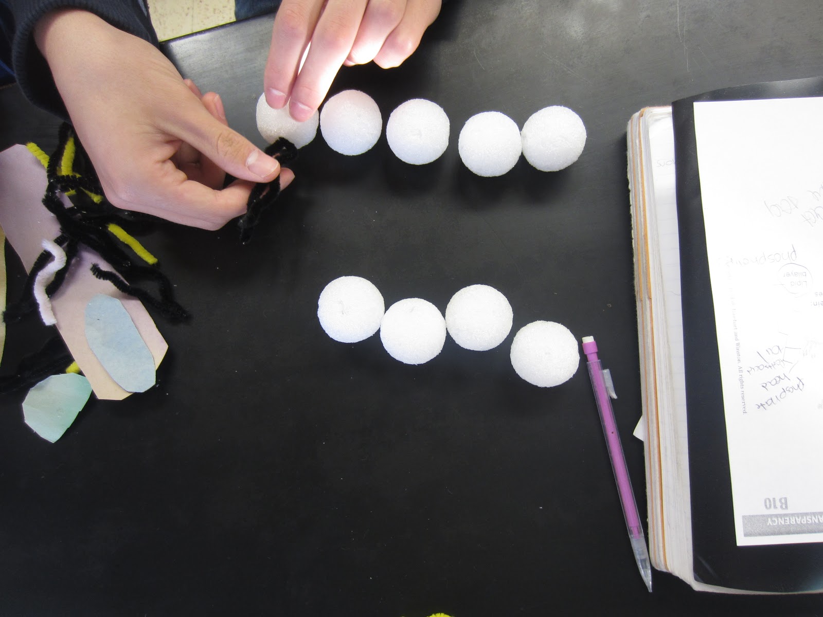
I then had you start building your cell membrane models.  This was a fairly "unguided" activity because I wanted you to look at your notes and using all the materials found in a bag - you were to create your own cell membrane AND be able to tell me all the parts/functions.  Here were some pics of how that went.  
Adding Non-polar fatty acid tails to the phosphate
heads
Phospholipid bilayer with some cholesterol
stuck in between the lipid tails (to help
stabilize the membrane)
Now to add the proteins - this one spans the entire membrane
which would allow molecules to move into and out of the cell.
Because of this, we could infer this is a transport protein

Completed cell membrane model!
After you built the models and described each part to me you answered and turned in the questions on the back of your cell membrane diagram (see below)  You ALSO added two questions to it.
1.  I have type A blood.  What part of the cell membrane surrounding my red blood cells determines that I have type A blood?

2.  TSH (Thyroid Stimulating hormone) binds to thyroid cells and initiates the production of T3 and T4 (Thyroid hormones), what part of the cell membrane does TSH bind to?


Tuesday, November 6, 2012

Cell membrane discussion


Daily Questions - November 6th
The cell membrane is made up of a bilayer made of two parts
1. Polar Phoshpate heads which are attracted to water (Hydrophilic)

2.  NonPolar Fatty Acid (lipid) Tails which are repelled by water (Hydrophobic)

Because of this, what kind of molecules can pass freely through the cell membrane? Small nonpolar molecules such as oxygen, carbon dioxide and nitrogen gasses.

Other substances (larger/polar molecules) move through membrane proteins known as  Transport Proteins (Channel and carrier proteins)

After the daily questions we went through the notes and followed along with the following diagram, adding these specific notes to it.


After going through the notes specifically on phospholipids, I had you watch the first couple minutes of the the following video relating the bilayer to Musk Ox.
Understanding how the cell membrane is 3 Dimensional can sometimes be very difficult.  I used the analogy of a wiffle ball.  I used just a basic pingpong ball as a visual for you to help you see that this is ALL around the cell - we are just looking at a cross section of it.
This of the cell as this pingpong ball - The cell membrane would be the plastic all around it

If we cut the the cell in half we would be looking at the inside
of the cell (where all the organelles and cytoplasm are)
Any of the images we are looking at of the cell membrane would be like
looking JUST at the plastic part that I colored black on here.